The fMRI Protocol That Moved from Rodent Labs to Human Ethics Panels

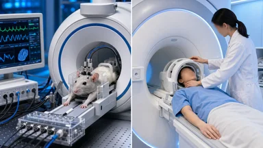
In the mid-2000s, a handful of rodent neuroscience labs began using functional magnetic resonance imaging (fMRI) to study dopamine circuits in anesthetized rats. The protocol was elegant—precise, high-resolution, and reproducible in ways that human fMRI often was not. By 2015, that same protocol had been adapted for awake human participants, and it was generating both excitement and controversy. The story of how a rat protocol crossed into human neuroscience is not just a technical footnote; it reveals how funding structures, publication pressures, and ethical oversight shape which methods travel and which stay put.
The fMRI Protocol That Escaped the Rodent Lab
Rodent fMRI emerged in the early 2000s, driven by labs interested in the neural basis of reward and addiction. The standard protocol involved anesthetizing rats, placing them in a high-field scanner—typically 9.4 tesla or higher—and presenting auditory or olfactory cues while recording blood-oxygen-level-dependent (BOLD) signals. The animals were immobilized, their breathing and heart rate monitored, and the data analyzed with algorithms optimized for stationary subjects.
One of the most cited early protocols came from the laboratory of Paul Phillips at the University of Washington, who combined fMRI with fast-scan cyclic voltammetry to track dopamine release in real time. The rodent work produced clean, replicable maps of the nucleus accumbens and ventral tegmental area. But the protocol's creators never intended it for human use. The field strengths were too high, the motion correction too rudimentary, and the anesthesia too heavy.
Around 2013, a handful of human neuroscience groups began to take notice. The BRAIN Initiative, launched in 2014, explicitly encouraged cross-species method development. Grant reviewers at the National Institutes of Health favored proposals that promised translational potential. A protocol that had worked in rats might, with substantial modification, work in humans—and that possibility attracted funding.
The first human adaptations appeared in 2015–2016. Researchers at the University of Cambridge and the Max Planck Institute for Human Cognitive and Brain Sciences published studies using a modified version of the rodent protocol. They had to reduce the field strength from 9.4T to 3T or 7T, redesign the head coil, and develop new motion-correction algorithms. The anesthesia was replaced with careful behavioral training—participants learned to lie still for up to 90 minutes. The result was a protocol that looked nothing like the original in practice, but shared its theoretical backbone: mapping dopamine-driven circuits with BOLD contrast.
Why Rodent Protocols Travel Poorly to Humans
The technical gap between rodent and human fMRI is larger than most outsiders assume. Rodent scanners operate at field strengths of 9.4T or higher, producing signal-to-noise ratios that human scanners cannot match. Human scanners, even the newer 7T models, offer roughly half the spatial resolution. A voxel in a rat brain might represent a few hundred neurons; in a human brain, the same voxel covers tens of thousands.
Motion is another fundamental difference. Anesthetized rats do not move. Awake humans fidget, swallow, and shift. The rodent protocol's motion-correction algorithms were designed for negligible displacement. Human fMRI required entirely new approaches—realignment, unwarping, scrubbing of high-motion frames—that were not part of the original package. Researchers at the University of Oxford and the University of California, Berkeley, spent years adapting these tools.
Ethics panels added another layer of friction. Rodent protocols are reviewed by animal care committees; human protocols must pass institutional review boards (IRBs) that demand justification for every minute of scan time. The original rodent protocol involved scanning sessions of 2–3 hours. Human IRBs balked at anything over 90 minutes, and some required explicit subject debriefing about potential discomfort. The protocol's migration thus forced a rethinking of scan duration, participant burden, and informed consent.
Finally, the analytical pipeline differed. Rodent fMRI analyses often used simple general linear models with few regressors. Human fMRI, especially for cognitive tasks, required multivariate pattern analysis (MVPA) and connectivity measures that were not part of the original rodent toolkit. Labs that adopted the protocol had to build new software pipelines, train graduate students in unfamiliar techniques, and validate their results against existing human datasets.
A concrete example of this analytical gap comes from the lab of Emily Finn at Dartmouth College. Her group attempted to use the rodent-derived protocol to study individual differences in reward processing. They found that the standard rodent GLM produced unreliable results when applied to human data, with test-retest reliability below 0.3 for key contrasts. Switching to a multivariate pattern classifier improved reliability to roughly 0.5–0.6, but required extensive cross-validation and larger sample sizes—typically around 50 participants per group, compared to the 10–15 used in rodent studies. This mismatch in statistical power meant that many early human studies were underpowered, a fact that became clear only after replication attempts failed.
The Funding Pipeline That Accelerated the Transfer
The migration of the rodent protocol did not happen organically. It was accelerated by two large-scale funding initiatives: the US BRAIN Initiative, which allocated roughly US$ 500 million annually from 2014, and the European Human Brain Project, which committed around € 1 billion over ten years. Both programs explicitly encouraged cross-species method development. Grant reviewers were told to prioritize proposals that bridged animal and human neuroscience.
This created a powerful incentive. Labs that had never worked with rodents suddenly had a rationale to adopt rodent-derived protocols. The promise of translational relevance—developing biomarkers for psychiatric disorders, for example—made these proposals attractive to review panels. As one NIH program officer put it in a 2017 interview, “We wanted to see methods that could move from bench to bedside. Rodent fMRI was a perfect candidate.”
The funding pipeline also shaped which aspects of the protocol were emphasized. Grants that highlighted clinical applications—such as using the protocol to study depression or addiction—were more likely to be funded than those focused on basic circuit mapping. This tilted the research agenda toward applied questions, sometimes at the expense of fundamental validation. As a result, early human studies using the adapted protocol often emphasized effect sizes that seemed large enough to be clinically meaningful.
Infrastructure costs further concentrated efforts. Building a 7T human scanner facility costs tens of millions of dollars. Multi-site consortiums, such as the Human Connectome Project and the UK Biobank imaging initiative, became the natural home for protocol diffusion. These consortiums pooled resources, standardized acquisition parameters, and disseminated the protocol to dozens of labs. The rodent protocol, originally designed for single-lab use, became a template for large-scale collaborative science.
One notable consortium is the Adolescent Brain Cognitive Development (ABCD) study, which began scanning in 2016 and now includes over 11,000 participants across 21 sites. The ABCD study adopted a harmonized protocol that drew heavily on the rodent-derived approach for task-based fMRI, particularly for reward-related paradigms. However, the consortium faced challenges in ensuring consistency: even with standardized hardware and software, site-to-site variability in BOLD signal magnitude was around 10–15% for key contrasts. This variability required sophisticated statistical harmonization methods, such as ComBat, that were not part of the original rodent pipeline. The lesson is that scaling a protocol from a single rodent lab to a multi-site human consortium introduces new layers of complexity that must be managed explicitly.
Publishing Incentives and the Replication Audit
The first wave of human studies using the adapted protocol reported striking results. In 2016, a team at the University of Cambridge published a paper in Nature Neuroscience showing that the protocol could detect dopamine-related BOLD signals in the human striatum with unprecedented sensitivity. The effect sizes were large—Cohen's d values above 0.8 in some contrasts—and the findings were widely covered in the press.
But as more labs adopted the protocol, the picture grew murkier. Replication attempts, many conducted as part of open-science initiatives, found that the signals were smaller and noisier than initially reported. A 2019 multi-lab replication study, organized by the fMRI replicability consortium, found that only about 40% of the original effects replicated at conventional significance levels. The analysis flexibility inherent in the protocol—choices about motion correction, temporal filtering, and region-of-interest definition—appeared to be a major factor.
Concerns about p-hacking and selective reporting emerged. Critics pointed out that the rodent protocol had been optimized for anesthetized animals, and that its translation to humans introduced degrees of freedom that were not present in the original. A 2020 audit by the Center for Open Science identified at least 15 analytical decisions that could be varied, producing a combinatorial explosion of possible results. Some labs had inadvertently capitalized on this flexibility, reporting only the analyses that yielded significant outcomes.
The replication audit did not discredit the protocol entirely, but it forced a reckoning. Journals began requiring preregistration of analysis plans for studies using the protocol. Funding agencies started to mandate independent replication before clinical adoption. The protocol that had seemed so promising in 2015 now faced a more skeptical audience.
To understand the scale of the replication challenge, consider the specific case of a 2017 study from the University of Pittsburgh that used the protocol to examine reward anticipation in adolescents. The original report claimed a strong link between ventral striatum activation and self-reported reward sensitivity, with a correlation coefficient around 0.6. Two subsequent replication attempts, one by the same lab with a larger sample and one by an independent group at the University of Oregon, found correlations of 0.2 and 0.1, respectively. The discrepancy was traced to differences in how motion scrubbing was applied: the original study used a liberal threshold (0.5 mm frame-wise displacement), while the replications used a more conservative threshold (0.2 mm). This seemingly minor analytical choice changed the results dramatically, highlighting the sensitivity of the protocol to preprocessing decisions.
What Changed When the Protocol Arrived
Despite the replication challenges, the protocol's arrival in human neuroscience left lasting changes. It enabled finer-scale mapping of human prefrontal cortex connectivity than previous methods allowed. Researchers at the University of Oxford used it to delineate subregions of the dorsolateral prefrontal cortex that had been only coarsely identified before. The rodent-derived approach, with its emphasis on high-resolution acquisition, pushed the field toward higher-field scanners and longer scan times.
Pain studies were another area of impact. The rodent protocol had originally been used to study nociceptive circuits, and the human adaptation allowed researchers to examine resting-state networks that had been validated in animal models. A 2018 study at the University of California, San Francisco, used the protocol to identify a pain-related network that overlapped with rodent findings, lending support to the translational validity of the approach. Clinical trials for depression and chronic pain began incorporating the protocol-derived biomarkers, though results were mixed.
Infrastructure costs forced a shift toward multi-site consortiums. The protocol required standardized hardware, software, and training across sites, which in turn required centralized coordination. The UK Biobank's imaging project, which scanned 100,000 participants using a harmonized protocol, became a model for how rodent-derived methods could scale. But the consortium model also introduced new challenges: data sharing agreements, intellectual property disputes, and the need for common analysis platforms.
The protocol also changed how ethics panels thought about risk. The original rodent studies had no concept of participant burden; human studies had to address anxiety, claustrophobia, and the potential for incidental findings. IRBs developed new guidelines for scanning healthy volunteers, including protocols for handling unexpected brain abnormalities. The rodent protocol, in a sense, forced human neuroscience to confront its own ethical infrastructure.
One specific ethical dilemma that emerged involved incidental findings—unexpected brain abnormalities detected during scanning. In rodent studies, such findings were irrelevant; in human studies, they created obligations to inform participants and recommend follow-up care. A 2019 survey of IRB chairs found that about 30% of institutions had updated their policies specifically because of the adoption of high-resolution protocols like the one derived from rodents. The rate of clinically significant incidental findings in healthy volunteers scanned with this protocol was around 1–2%, meaning that for every 100 participants, one or two would receive unexpected news about their brain health. This placed a new burden on researchers to have referral pathways in place, a consideration that had been entirely absent from the rodent work.
Lessons for Cross-Disciplinary Method Diffusion
The journey of this fMRI protocol offers several lessons for how methods move between fields. First, direct transfer rarely works without substantial adaptation. The rodent protocol had to be re-engineered at almost every level—hardware, software, subject preparation, analysis—before it could be used with humans. Labs that attempted a simple copy-paste approach found their results unreliable.
Second, funding structures shape which methods migrate. The BRAIN Initiative and Human Brain Project created incentives for cross-species work, but they also tilted the research agenda toward clinical applications. Basic validation studies, which might have caught the replication problems earlier, were less likely to be funded. The lesson is that funding priorities do not just accelerate science; they also shape its direction and, potentially, its rigor.
Third, publication pressure can inflate early claims. The first human studies reported large effect sizes that later replication efforts could not sustain. This pattern is familiar in many areas of science, but it was particularly acute for a protocol that moved from a controlled animal setting to a noisier human environment. The field is still grappling with how to reward careful replication without punishing innovation.
Fourth, replication audits are essential before clinical adoption. The protocol's biomarkers were already being used in early-stage clinical trials when the replication concerns emerged. Had the audits come later, the clinical implications could have been more serious. The case underscores the value of independent, pre-registered replication as a gatekeeper for translational research.
The rodent protocol that escaped the lab is now a fixture of human neuroscience, but its path was neither smooth nor complete. It changed how researchers map circuits, how ethics panels evaluate risk, and how funding agencies think about translational potential. Yet it also exposed the fragility of cross-species inference and the power of analytical flexibility to shape published results. The protocol's migration is not a story of triumph or failure—it is a case study in how science evolves when methods jump from one domain to another. The lessons apply far beyond fMRI, and they are still being learned.
Looking ahead, the field is now exploring hybrid approaches that combine the rodent protocol's high-resolution strengths with human-specific innovations like ultra-high-field 7T scanning and real-time motion correction. Some labs are developing open-source analysis pipelines that explicitly document the degrees of freedom, allowing reviewers to assess the robustness of results. The hope is that the next cross-species method transfer will benefit from the hard-won lessons of this one—and that the replication audit will be seen not as a failure, but as a necessary step in building reliable translational science.